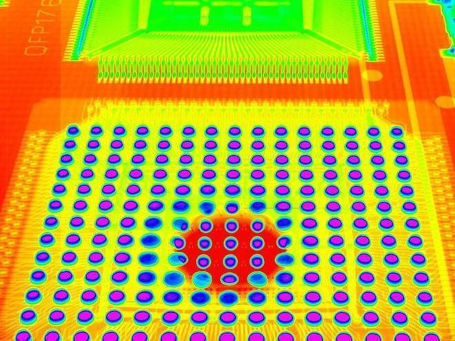
X-ray CT mainly consists of the following three parts
Release time:2024-01-31Publisher:Jeenoce
X-ray CT (ICT) is computer tomography or computed tomography imaging. Although the theoretical and mathematical theories related to tomography were first proposed by J Radon proposed, but it only became a new imaging technology after the emergence of computers and their integration with radiology. In the industrial sector, especially in the fields of non-destructive testing (NDT) and non-destructive evaluation (NDE), there are even more remarkable aspects.
X-ray CT refers to nuclear imaging technology applied in industry. The basic principle is based on the attenuation and absorption characteristics of radiation in the detected object. The ability of a substance to absorb radiation is related to its properties. So, using X-rays emitted by radioactive isotopes or other radiation sources with a certain energy and intensity, or γ The attenuation pattern and distribution of X-rays in the detected object may be displayed by detectors to obtain detailed information about the interior of the object, which can be displayed in the form of images using computer information processing and image reconstruction techniques.

1. The instrument should be placed in a clean and dry room, and efforts should be made to avoid surface contamination of optical parts, rusting of metal parts, and dust and debris falling into the moving guide rail, as this can affect the performance of the instrument.
2. After use, the working surface should be wiped clean at any time and can be covered with a dust cover.
3. The transmission mechanism and motion guide rail should be lubricated regularly to ensure smooth movement and maintain good working condition.
4. If the glass and paint surfaces on the workbench are dirty, they can be wiped clean with neutral cleaning agent and water.
5. All electrical connectors of the instrument should generally not be unplugged. If they have already been unplugged, we need to correctly plug them back in according to the markings and tighten the screws.
X-ray CT mainly consists of the following three parts:
The scanning section consists of an X-ray tube, detector, and scanning frame;
2. The computer system stores and calculates the information data collected through scanning;
3. Image display and storage system, which displays computer processed and reconstructed images on the TV screen or captures images using multiple cameras or laser cameras. The number of detectors has evolved from one to as many as 4800. The scanning methods have also evolved from translation/rotation, rotation/rotation, rotation/fixation to the recently developed spiral CT scanning. The computer has a large capacity and fast computation, which can achieve immediate image reconstruction. Due to the short scanning time, artifacts can be avoided. The scanning method used in high-speed CT scanning is different from the former. Micro CT scanning time can be as short as 40ms or less, and multiple frames of images can be obtained per second. Due to the short scanning time, movie images can be captured.

