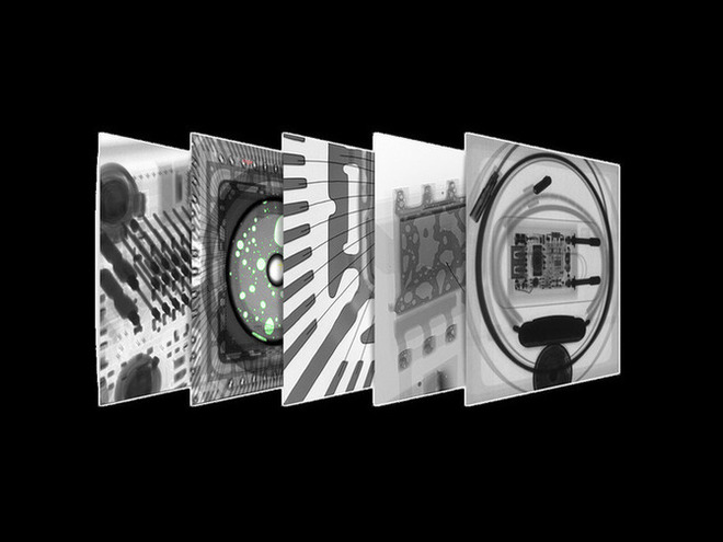
A Method for Improving the Image Quality of Microfocal X-ray Imaging
Release time:2023-11-24Publisher:Jeenoce
The use of microfocal X-ray for product inspection will affect the quality of the image after imaging, which will affect our judgment of defects in the tested product. The standards for judging image quality can be divided into subjective judgment and objective judgment. Subjective judgment is the direct judgment of the quality of an image through human vision and intuition; Objective judgment mainly relies on certain quantitative data to determine the quality of images. The signal-to-noise ratio is one of the important indicators for measuring image quality, and the higher the signal-to-noise ratio, the better the image quality.

There are several methods to improve the image quality of microfocal X-ray imaging:
Firstly, increase the tube current and increase the exposure time
The most typical denoising method for X-ray imaging process is to reduce CT imaging noise by increasing tube current and increasing exposure time. The generation of X-rays and their interaction with matter is a random process of photon counting, and increasing the tube current and exposure time can improve the signal-to-noise ratio of imaging. When the tube voltage is constant, the larger the tube current, the more X-ray photons are generated, and the higher the image signal-to-noise ratio. Increasing exposure time can be achieved by reducing the frame rate of image acquisition or by overlaying multiple frames of images. In addition, increasing the tube voltage of the microfocus X-ray source can enhance the ability of rays to penetrate the workpiece and improve image contrast.
Secondly, increase the amplification ratio
Compared to conventional focus X-ray sources, microfocal X-ray sources have a greater amplification ratio. By appropriately increasing the magnification ratio, the first step is to improve the detail resolution of the image. Secondly, the measured object can be kept away from the flat panel detector, and an increase in geometric distance can cause a portion of the scattered rays to decay in the air after penetrating the object, and the rate at which the scattered rays reach the detector will also decrease.
Thirdly, perform image fusion on multiple images
To address the image degradation caused by different focal sizes and shapes of micro focus X-ray sources in different radiation directions, multi angle image fusion can be used to process the images. The basic steps of this method are to capture an image of the workpiece at a certain angle, rotate the workpiece 90 degrees around the center of the beam, and then capture another image. Then, the two images are fused to achieve the best spatial resolution in both horizontal and vertical directions. Image fusion is an advanced image processing technique that integrates information from multiple source images. Its purpose is to inherit redundant and complementary information from multiple source images, in order to enhance the information in the image and increase the reliability of image understanding.

